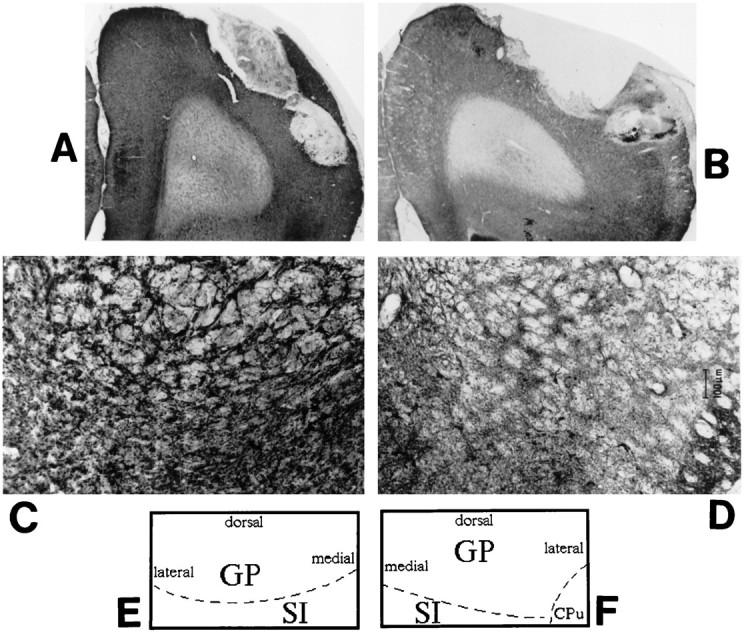
Fig. 5.

Representative coronal brain sections (stained for AChE) from a sham-lesioned animal (A) and a 192 IgG-saporin-lesioned animal (B) demonstrating representative placement of the microdialysis probe in the frontoparietal cortex. Again, the lesioned cortex shows a marked reduction in AChE-positive fiber staining. C andD show coronal sections through the basal forebrain area of representative control and lesioned animals, respectively. AChE-positive fiber staining is markedly reduced in the lesioned basal forebrain, implying a loss of cholinergic neurons in the basal forebrain after cortical infusion of 192 IgG-saporin. Eand F schematically illustrate the anatomical zones present in C and D. GP, Globus pallidus; SI, substantia innominata;CPu, caudate putamen. Magnification: A,B, 5×; C, D, 25×.
