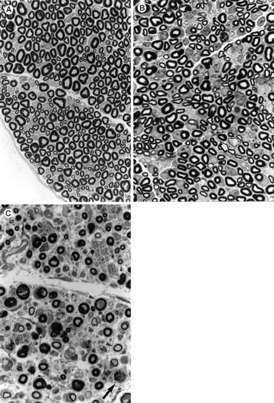
Fig. 2.

Light microscopy of an epon-embedded sciatic nerve stained with p-phenylenediamine (850×).A, Control. B, Section of a sciatic nerve from a V01 MDR3 transgenic mouse (11 d old) showing a decreased density of myelinated axons and several thinned myelin sheaths (arrow), compared with sections from control mice of the same age (not shown). C, Section of a sciatic nerve from a V01V01 MDR3 transgenic mouse, showing loss of myelinated nerve fibers, relatively thin myelin sheaths (arrowhead), and macrophage with myelin degradation products (arrow).
