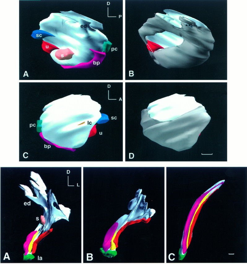
Fig. 3.

Top. Three-dimensional reconstruction of BMP4 and BMP7 gene expression of a stage 24 (E4) chick otocyst.A and B are medial views andC and D are lateral views of the same otocyst. This otocyst was reconstructed from a total of 44 horizontal, serial cryosections of 12 μm thickness. The most dorsal sections of the otocyst were missing, which resulted in a flattened image at the top. All odd-number sections were probed for BMP4, and all even-number sections were probed for BMP7. In A andC, all of the BMP4-positive, presumptive sensory organs are shown. Identification of each presumptive sensory organ was as described (Wu and Oh, 1996). The area for the presumptive lateral crista was under-represented in this specimen because of loss of adjacent sections for probing of BMP7 mRNA. In B andD, the BMP7-positive region of the otocyst is shown ingray. An anteromedial portion and a dorsolateral portion of the otocyst were negative for BMP7. All presumptive sensory organs were within a subset of the BMP7-positive region, except for a portion of the macula utriculi. The white dotted line demarcates the boundary of the presumptive macula utriculi area that overlapped with BMP7. The area in the endolymphatic apparatus that is marked byasterisks should be positive for BMP7. This gap in BMP7 expression was created by suboptimal alignment of sections in that area. Orientation: A, anterior; D, dorsal; P, posterior. bp, Basilar papilla; ed, endolymphatic apparatus; lc, lateral crista; s, macula sacculi; pc, posterior crista; sc, superior crista; u, macula utriculi. Scale bar, 100 μm.
