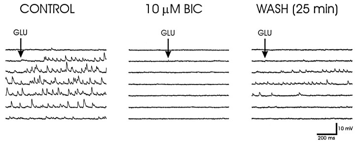
Fig. 2.
Glutamate-evoked IPSPs. Left, Glutamate microstimulation (GLU) at a position ventral to the fornix elicited reversed IPSPs in a PVN parvocellular neuron recorded with a KCl-filled microelectrode.Middle, Bath application of the GABAA-receptor antagonist bicuculline methiodide (BIC) for 15 min completely blocked the effect of glutamate microapplication at the same site and with the same application parameters. Right, Partial recovery of the glutamate-evoked IPSPs was seen after 25 min of washout of the bicuculline. The membrane potential was held at −105 mV with negative current injection.

