
Fig. 6.
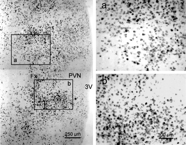
Distribution of GAD65 mRNA-containing neurons in the horizontal plane of the hypothalamus. Low-magnification photomontage of a horizontal section labeled with the GAD65 riboprobe (left). GAD65 mRNA is expressed in neurons scattered throughout the hypothalamus, including cells surrounding the PVN, which itself is relatively devoid of GAD65 mRNA-containing cells. The sites rostral and caudal to the fornix that were stimulated by glutamate microdrops to elicit IPSPs in PVN neurons are represented byboxes a and b, respectively, which are shown at higher magnification to the right. The midline is to the right, and rostral is up in each photomicrograph. The calibration in b pertains to both a and b. Fx, Fornix;3V, third ventricle.