
Fig. 5.
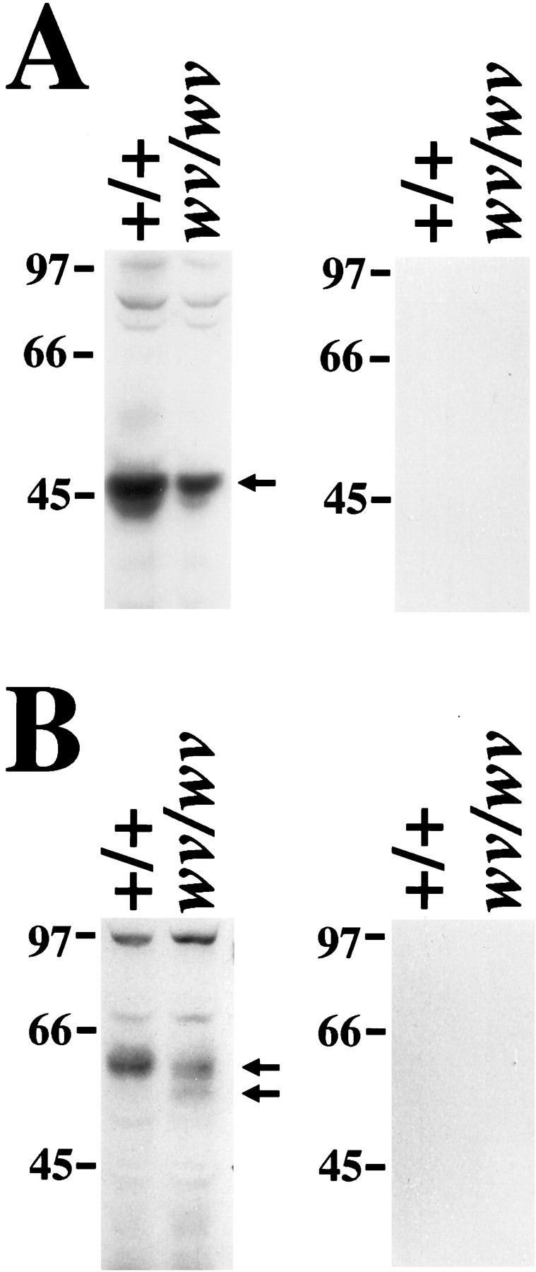
Decreased level of both GIRK2 and GIRK1 proteins in the wv brain. A, Left, Western blotting with antibody against GIRK2 shows that there is less GIRK2 protein in the wv brain than in wild-type brain. Fifty micrograms of membrane from wild-type littermate orwv brain were loaded onto each lane. B,Left, Western blotting with antibody against the N terminus of GIRK1 demonstrates that there is a slight decrease in the 58–60 kDa GIRK1 band, but there is an additional 55 kDa band, which may represent an unglycosylated form of GIRK1 in the wvbrain. A, B, Right, Peptide competition controls for antibodies against the N terminus of GIRK2 or GIRK1, respectively.