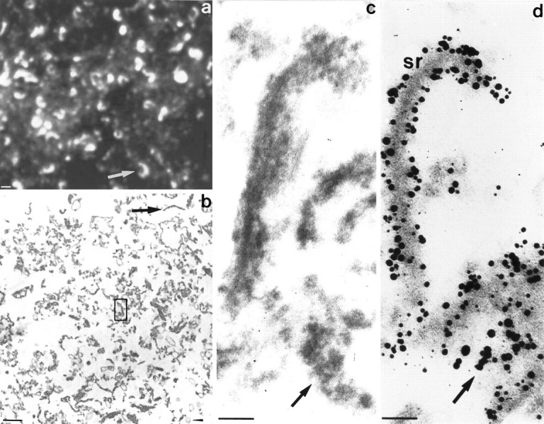
Fig. 7.

a–d, Characterization of the SR-fraction. a shows a cryostat section of the SR-fraction labeled with the ribbon antiserum and processed for immunofluorescence and confocal laser scanning microscopy. Note the high density of horseshoe-shaped synaptic ribbons present in this fraction. b represents a conventional transmission electron micrograph of the SR-fraction. Note the presence of many bar-like ribbon profiles (arrow) surrounded by electron-dense material. Only a few contaminating membranous structures were present in this fraction (arrowhead).c is a higher, enlarged transmission electron micrograph similar to the one labeled in b (box inb). Note the bar-shaped ribbon profile surrounded by electron-dense material. The isolated synaptic ribbons appear more diffuse than ribbons in situ. In d, the SR-fraction was immunolabeled with the ribbon antiserum to analyze which components of the SR-fraction represent ribbon material. Both ribbons as well as the surrounding electron-dense material are immunolabeled with the ribbon antiserum, indicating that the dense material represents a disassembly product of synaptic ribbons. Scale bars: a, b, 1 μm; c, d, 100 nm.
