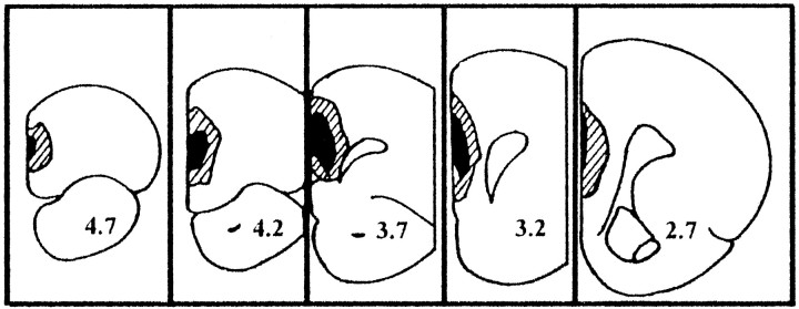
Fig. 1.
Schematic drawing of coronal sections (modified from Paxinos and Watson, 1986) illustrating lesion boundaries and the areas of neural loss and gliosis determined from Nissl-stained coronal sections from PD60 rats with ibotenic acid lesion of the neonatal MPFC. The stippled lines and solid black areas indicate the largest and smallest lesions, respectively. Coordinates refer to distance in millimeters anterior to bregma (Paxinos and Watson, 1986).

