
Fig. 2.
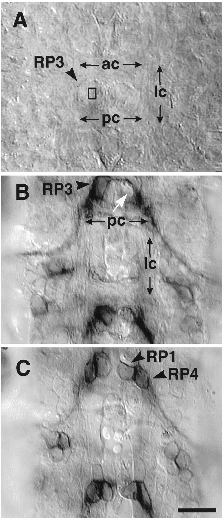
Laser ablation of RP3 in live stage late 15/early 16 embryos. A, The cell body of RP3 is imaged through the ventral surface of the dechorionated embryo. RP3’s cell body is located near the dorsal (internal) surface of the ventral nerve cord. Two RP3 cell bodies (arrowhead) can clearly be seen flanking the midline and bordered by the lateral commissures (lc) on the left and right, and above and below by the anterior and posterior commissures (ac,pc). The rectangle indicates the ∼1 × 2 μm target site of the laser. Every visible RP3 on one side was targeted. The contralateral RP3 seconds were left intact as internal controls. B, C, To test the specificity of the laser ablation with respect to neighboring cells, operated embryos were acutely filleted and stained with anti-Fasciclin III, which labels a subset of neurons, including RP1,RP3, RP4. B, In this segment seen from the dorsal view, there is no staining in the location of RP3 (white arrow), whereas the contralateral RP3 is still immunopositive for FasIII. C, At the dorsal surface of the CNS, one cell layer above RP3, both RP1and RP4 can be seen on both sides. Scale bar, 10 μm.