
Fig. 5.
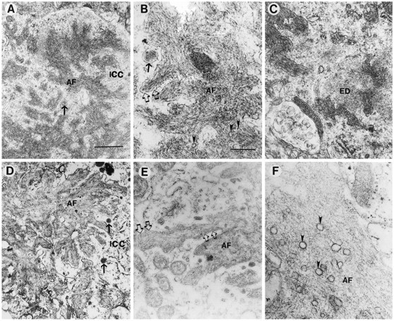
Comparison of the amyloid deposits in the PDAPP tg model and AD. Electron micrographs were obtained from the hippocampal region in the PDAPP tg mouse and from the frontal cortex in AD. Low-power (A) and high-power (B) view of amyloid fibrils (AF) in the PDAPP tg mouse showing dense deposits surrounded by a membrane (open arrows) and the neuronal intracytoplasmic compartment (ICC) containing electrodense granular material and dense-core neurosecretory vesicles (arrow). Occasional poorly defined clear vesicles (arrowheads) were associated with the amyloid fibrils in PDAPP mouse plaques.C, In the PDAPP tg mouse, the cytoplasmic component of neurons close to the extracellular amyloid fibrils (AF) showed the presence of amorphous electrodense material (ED). Low-power (D) and high-power (E) view of the amyloid deposits in AD showed abundant dense fibrils (AF) surrounded by a membrane (open arrows) and a cytoplasmic compartment (ICC) containing dense-core neurosecretory vesicles (arrows) and electrodense granular material.F, Clear vesicles were prominently associated with the amyloid fibrils (AF) in AD plaques (arrowheads). Scale bars: A, 1 μm;B, 100 nm.