
Fig. 6.
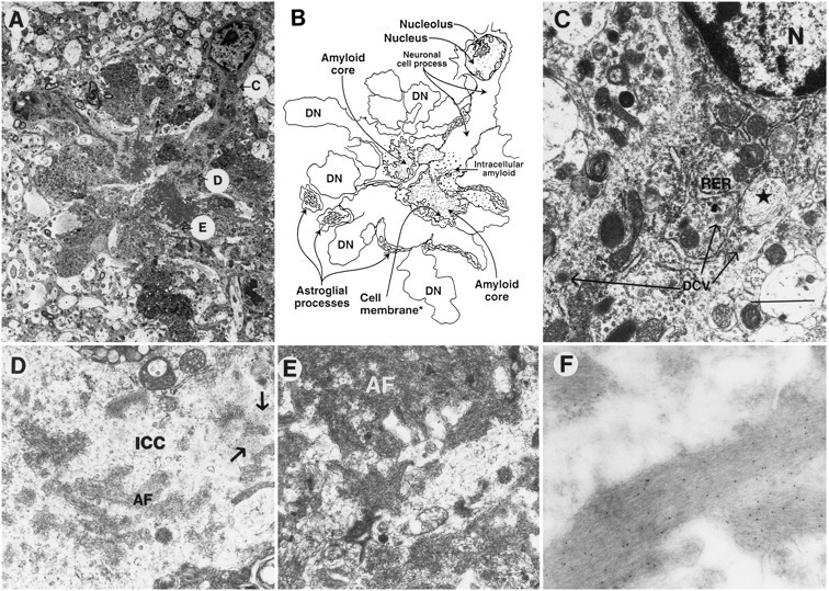
Subcellular neuronal alterations associated with the neuritic plaques in PDAPP tg mouse. Electron micrographs were taken from a lesion in the hippocampus. A, Low-power view of a neuritic plaque composed of a prominent neuronal element accompanied by extensive neuritic dystrophy, amyloid deposits, and astroglial cell reaction. The letters inside circles correspond to subsequent panels that identify various characteristics of the neuronal element in the plaque in greater detail. B, Diagrammatic representation of the neuritic plaque illustrated in A.C, Proximal to the nucleus (and distal to the amyloid core), the neuron displayed abundant RER, neurosecretory dense-core vesicles (long arrows), and peculiar elliptical bodies containing fibrilo-tubular material (*). Scale bar, 3 μm.D, Distal to the cell nucleus (and proximal to the amyloid core), the intracellular cytoplasmic compartments (ICC) of the neuronal process displayed amyloid fibrils (AF) and amorphous electrodense granular material (arrows) surrounded by a membrane-forming digitating processes (E). The neuronal cytoplasm surrounding the extracellular amyloid fibrils (AF) showed diffuse granular material and mitochondria. F, Immunogold labeling with a monoclonal antibody against β-amyloid (3D6) showed amyloid fibrils decorated with the 5 nm gold particles.