
Fig. 7.
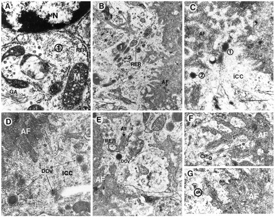
Additional neuronal subcellular characteristics associated with amyloid formation in the PDAPP tg mouse. Electron micrographs were obtained from the hippocampus and are derived from the neuritic plaque presented at the bottom of Figure 3D.A, Proximal to the nucleus (N) (and distal to the amyloid core), the neuron displayed, in addition to RER and elliptical inclusion bodies (1), a prominent Golgi apparatus (GA) and mitochondria (M). B, Distal to the cell nucleus (and proximal to the amyloid core), the cytoplasm of the neuronal process displayed the presence of compact electrodense organelles surrounded by a membrane (1) adjacent to RER, mitochondria, and neurosecretory vesicles (2).C, D, Dense-core neurosecretory vesicles (1, 2) were abundant in the intracytoplasmic compartment (ICC) of the neuron adjacent to the extracellular amyloid fibrils (AF). E, Intracellular amyloid fibrils (AF, black letterhead) adjacent to RER were present in the cytoplasmic compartment of the neurons near the extracellular amyloid fibrils (AF, white letterhead). Dense-core neurosecretory vesicles (DCV) were also present. F,G, Coated pits (CP) in the cytoplasmic compartment of the neurons were closely associated with the plasma membrane (circle) surrounding extracellular amyloid deposits (AF). Scale bar, 400 nm.