
Fig. 9.
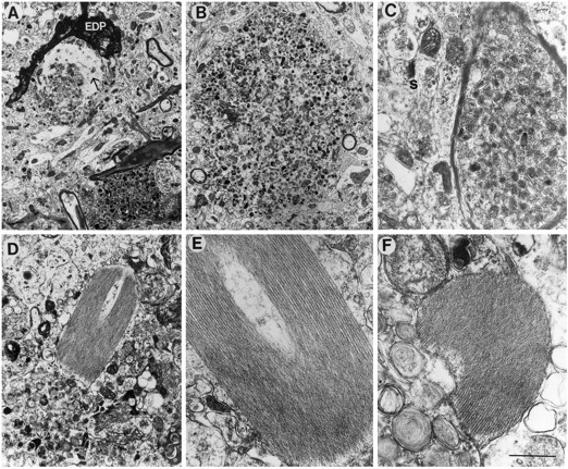
Neuritic alterations in PDAPP tg mice. Electron micrographs were obtained from the hippocampus. In PDAPP tg mice, the dystrophic unmyelinated (A, B) and myelinated neurites (C) contained abundant electrodense laminar and multivesicular bodies. Some neurites contained fine filaments (10 nm; arrow) and were surrounded by electrodense processes (EDP). Other neurites contained characteristic crystals displaying an array of symmetrically organized tubules (D–F). Scale bar, 2 μm.