
Fig. 2.
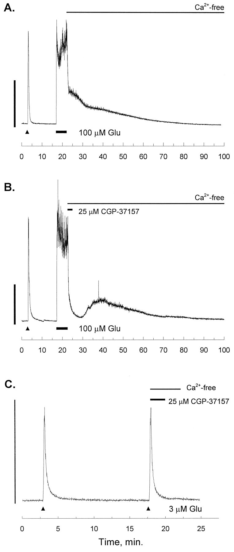
[Ca2+]i recordings after intense glutamate stimulation. In each trace, the small triangle indicates a 15 sec stimulus of 3 μmglutamate (Glu) and 1 μm glycine (gly) as an internal control (White and Reynolds, 1995). The experimental stimulus is applied after a 15 min recovery, and in each trace, the second Glu stimulus is washed out with a nominally Ca2+-free solution (no EGTA). A, After recovery from the internal control, 100 μm Glu and 1 μm gly were applied for 5 min (at theline), and the Glu was followed immediately by a nominally Ca2+-free solution (no EGTA). The trace is representative of 13 experiments. B, After recovery from the internal control, 100 μm Glu and 1 μmgly were applied for 5 min (at the line), and the Glu was followed immediately by a solution of the mitochondrial Na+/Ca2+-exchange inhibitor CGP-37157 (25 μm) in Ca2+-free buffer. After 2 min the CGP-37157 was washed out with Ca2+ -free buffer. The trace is representative of seven experiments.C, After recovery from the internal control, a second identical Glu pulse (15 sec, 3 μm Glu and 1 μm gly) was applied (at the small triangle), and the Glu was followed immediately by a Ca2+-free solution of CGP-37157 (25 μm). After 2 min, the CGP-37157 was washed out with Ca2+-free buffer for an additional 2 min before bath application of ordinary HBSS. The trace is representative of six experiments. In each panel, the scale bar indicates 1 μm[Ca2+]i.