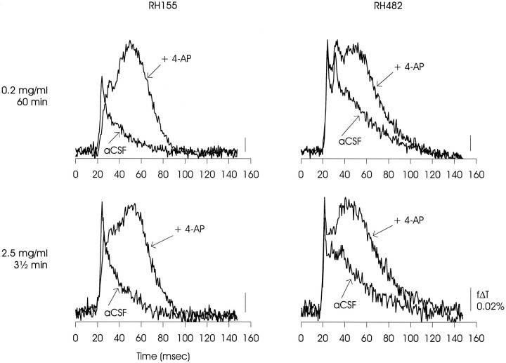
Fig. 1.
Matrix of optical signals from area CA1 of rat hippocampal slice showing that similar differences in voltage-dependent signals obtained in control and 4-AP-containing solutions were observed using either of two related voltage-sensitive dyes, RH-155 or RH-482, and either of two different loading protocols, 0.2 mg/ml for 60 min or 2.5 mg/ml for 3 1/2 min. The variation between RH-155 and RH-482 evident in the ACSF relaxations, particularly at the lower staining concentration, was within the range observed in these experiments, and therefore separation of signals originating in neural or glial membranes may not be possible in this preparation (Konnerth et al., 1987). The data presented are representative of one experiment with RH-155 at 0.2 mg/ml, five experiments with RH-155 at 2.5 mg/ml, three experiments with RH-482 at 0.2 mg/ml, and two experiments with RH-482 at 2.5 mg/ml.

