
Fig. 2.
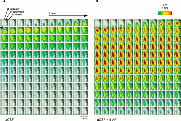
Propagation of depolarization through area CA1 of rat hippocampal slice before (A) and during (B) application of 4-AP (40 μm), illustrating potentiation of the initial voltage transient (EPSP/AP complex) and induction of a delayed depolarization by 4-AP (see Fig.4). The slice was stained with RH-155 and imaged at 0.6 msec/frame as described in Materials and Methods. The major layers of the slice are labeled in the first panel. The depolarization was measured as the fractional change in transmittance (f Δ T) in each pixel; this value is encoded inpseudocolor as indicated in the scale and is superimposed on a transmitted light image of the slice. The Schaffer collateral input from CA3 to CA1 was stimulated using bipolar tungsten electrodes positioned just below the lower edge of the image (black arrowhead), and in each sequence the stimulus was delivered between the first and second frames. The two finger-like projections in the top right portion of each image are from an electrode used for antidromic stimulation. The images inB were acquired after the slice had been exposed to 4-AP for 20 min; in general, we allowed 15–30 min for the effects of 4-AP to reach a steady state.