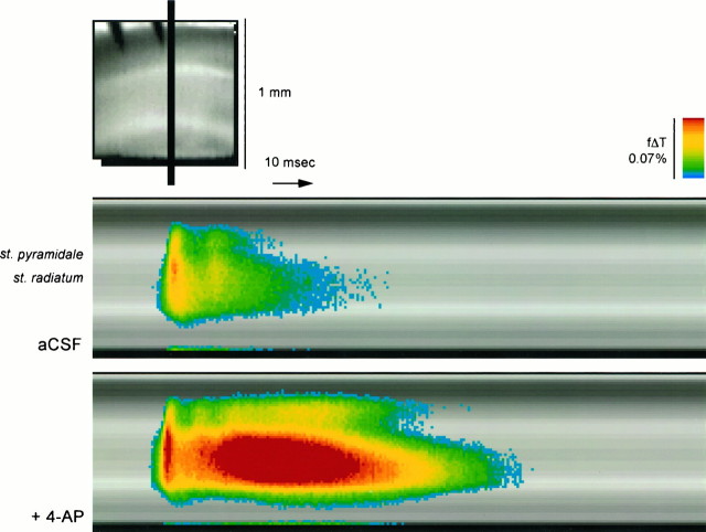
Fig. 3.
Time course of voltage signals in ACSF and in the presence of 4-AP. Voltage changes in the one-dimensional stripe indicated in the top panel were extracted from each frame and arranged in a time series in the bottom panels. Note the prominence of the 4-AP-induced delayed depolarization in the st. radiatum and its bidirectional lateral expansion.

