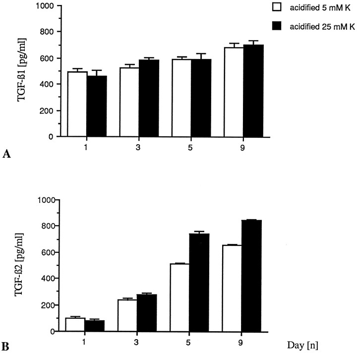
Fig. 8.
Cerebellar granule neurons maintained in either high or low K+ medium secrete latent TGF-β1 and TGF-β2. Cell-free conditioned media were transiently acidified as described in Materials and Methods, and both activated and nonactivated media were tested in triplicate for TGF-β1 and TGF-β2 in a quantitative sandwich enzyme immunoassay at the respective dilutions of 1:7 and 1:4. The detection limit of both immunoassays does not allow quantification of concentrations <60 pg/ml. A, Neurons grown in either low or high K+ concentrations produce equal amounts of latent TGF-β1, mostly in the first 24 hr of culture, with a slight increase over time in vitro. Naturally active TGF-β1 was not detected by the present technique. B, Neurons grown in either low or high K+ culture conditions release latent TGF-β2 with a continuous production over time in culture and a major contribution by high K+neurons. Naturally active TGF-β2 also was not detected.

