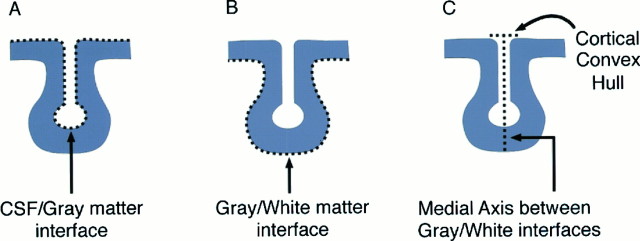
Fig. 1.
Rules for delineating sulci. The ability to resolve neuroanatomic boundaries is critical for accurate structure delineation. Three methods are shown for defining the interior course of sulci in cryosection images. The densitometric gradient afforded by 24-bit full-color images provides excellent color pigment differentiation and texture contrast at the exterior surface of the cortical laminae (A) and at gray-white matter interfaces flanking the sulci (B). Nevertheless, a medial axis definition (C), adopted here, provides a fundamental laminar path into the brain for each primary sulcus, the structural integrity of which is not compromised in regions where secondary sulci branch away, or at points of confluence with other sulci. In addition, methodC is adaptable for use with other anatomic imaging modalities such as MRI, in which cellular interfaces are blurred out or more diffusely represented. The course of medial axis is not affected by any purely symmetrical errors, which occur in identifying the opposing sulcal banks. It can therefore be identified in an accurate and reproducible way, even in low-contrast imaging modalities.

