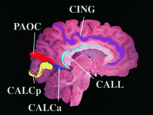
Fig. 2.
Sagittal projection of the full set of sulcal contours traced in the left hemisphere of a single brain. These sets of contours were derived from the full series of sectional images spanning the left hemisphere of one brain specimen. Orthogonally projected contours of the anterior and posterior rami of the calcarine sulcus (CALCa and CALCp), as well as the cingulate (CING), supracallosal (CALL), and parieto-occipital (PAOC) sulci, are shown overlaid on one representative sagittal section.

