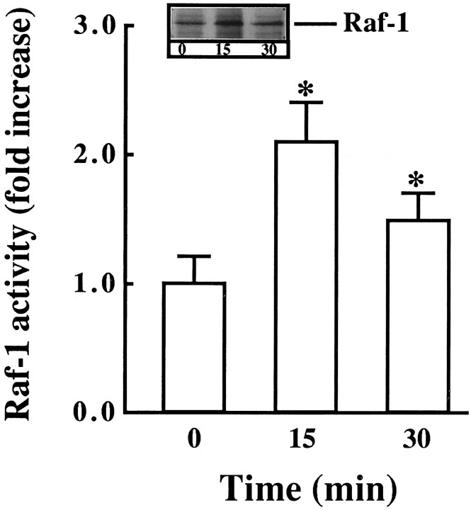
Fig. 7.
Co-precipitation of Raf-1 with Ras. Neuronal cultures were stimulated with 100 nm Ang II for the indicated times (min). Cell lysates were immunoprecipitated with anti-Ras antibody. Western blots of the immunoprecipitates were then probed with anti-Raf-1 antibody. Top, Representative autoradiogram. Bottom, Bands corresponding to Raf-1 were quantitated, and data from three immunoblots were presented as mean ± SE. Asterisks indicate significantly different from the zero time density.

