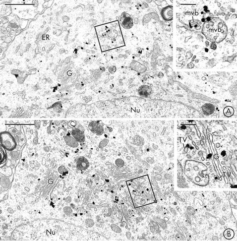
Fig. 1.

VMAT2 is localized to saccules of Golgi and multivesicular bodies in neuronal perikarya in the SN and VTA.A, Immunogold particles for VMAT2 are seen in the region of the Golgi apparatus (G) of a labeled perikaryon in the SNC, but are not detected in the adjacent rough endoplasmic reticulum (ER). The inset shows the boxed regionat higher magnification. Immunogold labeling for VMAT2 is seen along the limiting membranes of two multivesicular bodies (mvb).B, Immunogold labeling for VMAT2 is localized to the Golgi apparatus (G) of a perikaryon in the VTA. Many of the gold particles contact lateral saccules of Golgi lamellae (curved arrows). The inset shows the boxed region at higher magnification. Gold particles are localized to saccules of Golgi (G) and associated vesicles and tubulovesicles (TV). Nu, Nucleus. Scale bars: A, B, 1 μm; inset in A, B, 0.2 μm.
