
Fig. 6.
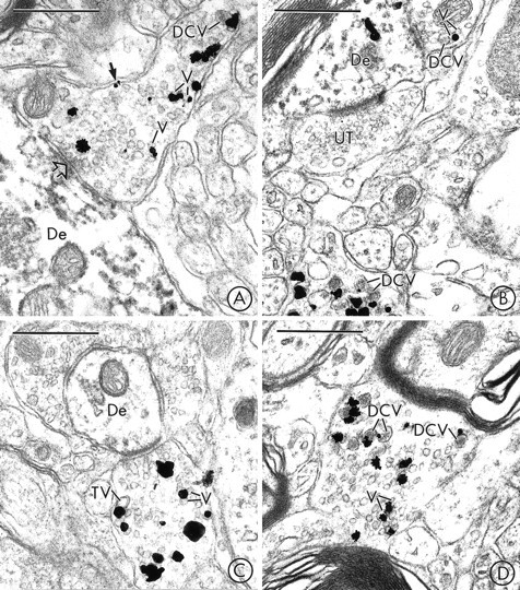
In the SN and VTA, VMAT2 is localized to SSVs and DCVs in axon terminals that usually lack detectable TH-immunoreactivity. A, Immunogold particles for VMAT2 are localized to small synaptic vesicles (V), an isolated DCV, and the plasma membrane (closed arrow) of an axon terminal in the SN that lacks detectable peroxidase reaction product for TH. The VMAT2-containing terminal forms a synaptic contact (open arrow) with a dendrite (De) that contains intense peroxidase reaction product for TH, but no detectable immunogold labeling for VMAT2. B, Immunogold particles for VMAT2 are seen in two axon terminals in the SN. The terminal at thetop contacts a dendrite (De) that is labeled with immunoperoxidase for TH and immunogold for VMAT2. The terminal at thebottom contains numerous VMAT2-labeled large dense-core vesicles (DCV). The VMAT2-immunoreactive terminals contain no detectable peroxidase labeling for TH. UT, Unlabeled terminal. C, Immunogold particles for VMAT2 are localized to small synaptic vesicles (V) and a larger electron-lucent tubulovesicular structure (TV) in an axon terminal in the VTA that lacks detectable peroxidase reaction product for TH. The VMAT2-labeled terminal contacts a dendrite (De) that is lightly immunoperoxidase-labeled for TH. D, An axon terminal in the VTA contains immunogold particles that are localized to the membranes of large dense-core vesicles (DCV) and small synaptic vesicles (V). The VMAT2-labeled terminal contains no detectable peroxidase reaction product for TH. Scale bars:A–D, 0.5 μm.