
Fig. 5.
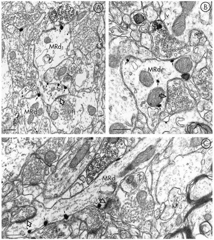
Electron micrographs in A–C show dendrites containing immunogold-labeling for MOR (MRd) that are apposed to terminals containing peroxidase labeling for LE (Et). The dendrite labeled for MOR-LI in A has one gold–silver particle (top, small arrow) oppositeEt, whereas the majority of particles (small arrows) are located along the plasmalemma of the head and neck of the spine (s) emerging from MRd. This spine receives an asymmetric synapse (open arrow) from an unlabeled terminal (ut). Each of the gold particles in the nearby dendrite and spine are also in contact with plasma membranes.B shows immunogold–silver labeling for MOR associated with both the plasma membrane and cytoplasmic organelles (small arrows) within a dendrite (MRd). MRd is apposed to a terminal showing immunoperoxidase label for LE (Et). In this terminal, the peroxidase is intensely localized to a large vesicle (lv). MRd also receives a symmetric synapse (open arrow) from an unlabeled terminal (ut). In C, a longitudinally sectioned dendrite (MRd) shows gold–silver immunolabeling for MOR (small arrows) localized along the plasma membrane, distal to Et. MRd is also postsynaptic to an unlabeled terminal (ut1) where the synaptic specialization (open arrow) appears asymmetric. The peroxidase-labeled Et can be compared with a second unlabeled terminal (ut2) in the same field. Scale bars, 0.3 μm.