
Fig. 10.
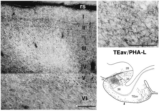
Dark-field photomicrograph of a dense core region in area 36, which is illustrated at the bottom right within the circumscribed area (same as Fig. 9A). The high-magnification photomicrograph of terminals with boutons (bright-field) at the top right is taken from layer III of the photomicrograph shown at the left. Scale bars:left, 0.25 mm; right, 0.1 mm.