
Fig. 4.
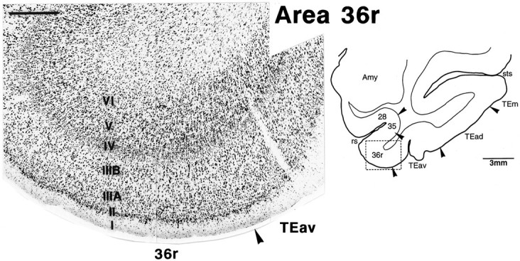
Cytoarchitecture of TEav and the rostral part of area 36 (36r) in a Nissl-stained section. The position of the photomicrograph is indicated by the box in the lower-magnification line drawing of the section on theright. The subdivision of layer III into IIIA andIIIB is clearer in 36r than in 36c (Fig. 3), and layer II is more distinctive with many darkly stained neurons and satellite glial cells in 36r. Such distinction is not clear in TEav. Scale bar, 0.5 mm.