
Fig. 5.
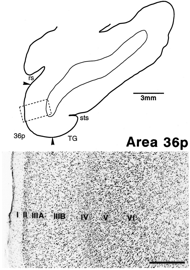
Cytoarchitecture of the polar part of area 36 (36p) in a Nissl-stained section. The position of the photomicrograph is indicated by the box in the line drawing of the section at the top. The distinction between IIIA andIIIB, and that between V and VI, is not clear. Also, layer IV is less distinctive than those in 36c and 36r. Scale bar, 0.5 mm.