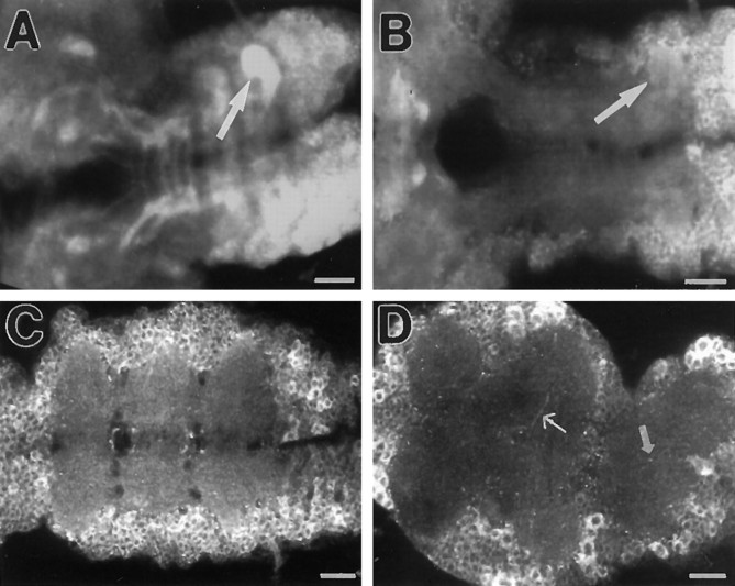
Fig. 6.
APPL immunoreactivity in the ventral ganglion during metamorphosis. Optical section through whole-mount preparations of ventral ganglions dissected from Canton S pupae at 0 hr (A), 6 hr (B), 12 hr (C), and 48 hr (D) after pupariation and stained with anti-APPL antibody. Signal in the thoracic neuromere (arrow in A andB) decreases after 6 hr, and by 12 hr APPL is distributed evenly in the neuropil of the ventral ganglion (C). By 48 hr, the level of APPL in the neuropil is low, and distinct processes (thin arrow) and varicosities (small arrow) are revealed. For A–D, anterior is to the left. Scale bars: A–D, 25 μm.

