
Fig. 7.
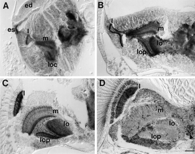
APPL immunoreactivity in the optic lobe during metamorphosis. Horizontal paraffin section of heads of 0 hr (A), 25 hr (B), and 60 hr (C) pupae and adult (D) stained with anti-APPL antibody. A, 0 hr after pupariation, APPL-immunoreactive photoreceptor axons project from the eye disk (ed) through the eye stalk (es) into the optic lobe. The lamina (l), the outer and inner medulla neuropils (m), and the two neuropils in the lobula complex (loc) in the optic lobes are highly stained with APPL. B, By 25 hr after pupariation, APPL immunoreactivity in the medulla neuropil starts to split into three layers, the two outer ones being connected by processes. In the lobula complex, the lobula (lo) and lobula plate (lop) neuropils are intensely stained. C, 60 hr after pupariation, the optic neuropils have rotated. In the lamina (l), axons are distinguishable. In the medulla (m), APPL is arranged in three layers, showing a columnar organization. D, In adult, intense APPL signal remains only in the lamina neuropil (l). In the medulla, isolated processes are APPL-im-munoreactive (thin arrows). Some cell bodies show higher APPL immunoreactivity (arrowhead; lamina cortex). For A–D, anterior is at the top and lateral at the left. Same magnification was used for all. Scale bar (shown in D): 25 μm.