
Fig. 1.
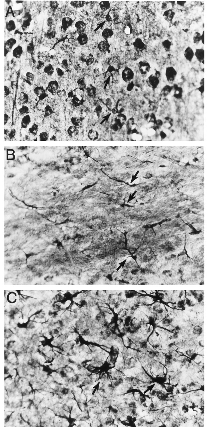
Cajal’s gold chloride sublimate technique to identify normal and reactive astrocytes. Some astrocytes in all three frames are indicated by arrows. A, Protoplasmic astrocytes in the gray matter of the uninjured cortex. The darkly stained round structures are neuronal soma. To visualize normal protoplasmic astrocytes in the cortex, it is necessary to overstain brain sections, and this results in the neuronal cell bodies being darkly labeled. In contrast, after injury, astrocytes can be easily revealed without the need for overstaining (e.g., in frameC; thus, neuronal cell soma in C is not prominent). B, Fibrous astrocytes in the uninjured corpus callosum. C, Reactive astrocytes in the cortex after traumatic injury. Magnification, 400×. It has been our experience that the Cajal method, compared with GFAP/Mac-1 immunofluorescence or GFAP immunoperoxidase-AEC methods, amplifies the differences in depth of field; this contributes to some of the cells appearing out of focus in this figure.