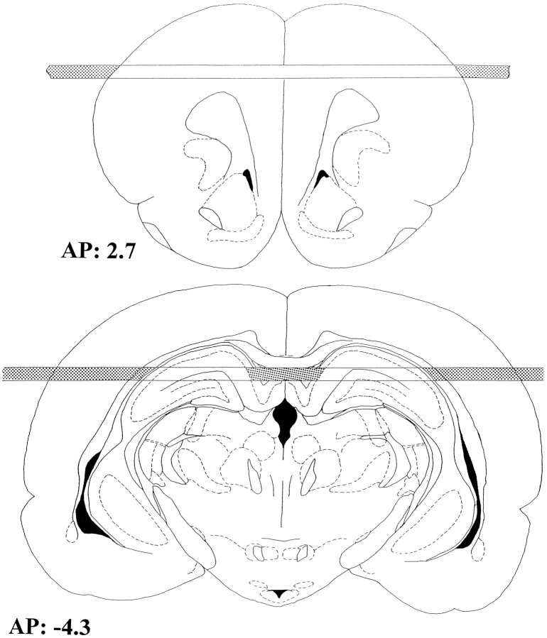
Fig. 1.
Schematic representation of the location of the microdialysis probes redrawn from Paxinos and Watson (1986).Shaded areas of the membranes represent the parts covered with epoxy glue. AP, 2.7: frontal cortex (top). AP, −4.3: hippocampus (bottom).

