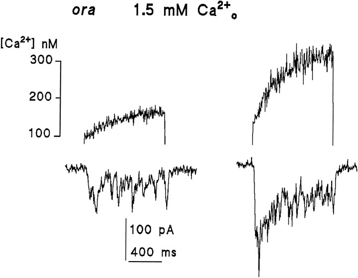
Fig. 6.
Ca2+ influx measured in real time in ora photoreceptors containing residual levels of rhodopsin. UV measuring flashes of 1 sec duration delivered toora (or ninaE) photoreceptors sometimes elicited small responses (lower traces) attributable to residual levels of rhodopsin, thus allowing direct measurement of Ca2+ influx (upper traces) during weak effective illumination. Traces from two different cells are shown using different intensities, generating responses of ∼20–40 pA (left) and 200 pA (right). Quantum bump noise can be clearly resolved in these small responses: as reported previously (Johnson and Pak, 1986), quantum bumps in ninaE mutants with greatly reduced rhodopsin levels were in fact typically larger than in WT. Substantial Ca2+ rises were detected in each case. The data from these and three other cells are plotted on Figure5b and show reasonable agreement with measurements made in WT photoreceptors using the two-flash paradigm.

