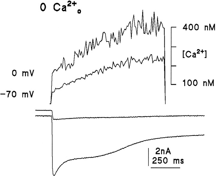
Fig. 9.
Light-induced Ca2+ signals measured in the absence of extracellular Ca2+. Substantial Ca2+ increases were detected in every cell in response to saturating UV-measuring stimuli: Ca2+ signals (upper traces), simultaneously recorded whole-cell currents at 0 and −70 mV (lower traces) (two different cells). The rise was at least as large in cells clamped at 0 mV as in those clamped at −70 mV, arguing against influx of residual Ca2+ as an explanation. Note also that the initial (dark) resting level of Ca2+ was higher in the cell clamped at 0 mV (see also Fig. 11). Bath contained 0 Ca2+, 2 mm EGTA, and 120 mmNaCl.

