
Fig. 4.
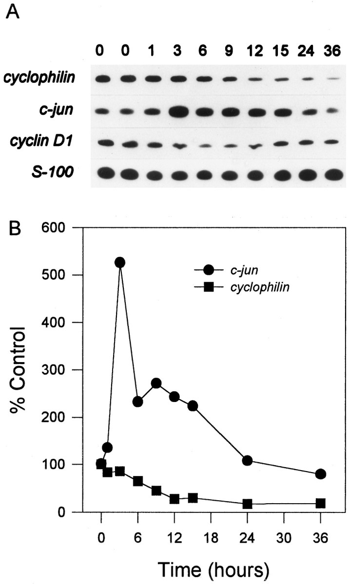
Analysis of mRNA levels in cerebellar granule cells. Cultures were switched to K5 − S, and cDNA was prepared from granule cells after 1, 3, 6, 9, 12, 15, 24, or 36 hr. cDNA from ∼4000 cells was used in a 50 μl PCR reaction, as described in Materials and Methods. Each transcript was analyzed from at least two different neuronal preparations. A, Representative response of cerebellar granule cells in K5 − S medium (comprehensive list in Table 1). cyclophilin was typical of genes that decreased. cyclin D1 decreased slightly and then increased to control levels by 36 hr. c-junincreased approximately fivefold during PCD. S-100, a marker for non-neuronal cells, remained relatively constant. B, Quantitation by PhosphorImager analysis of mRNA levels in cerebellar granule cells in K5 − S medium.