
Fig. 1.
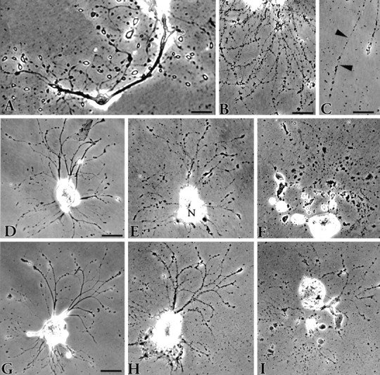
Phase-contrast views depicting the fate ofAplysia neurons in vitro.A, Neuritic processes 5 d after plating showing the bulbous, birefringent endings that have replaced the growth cones on the newly formed neurites. The appearance of these structures marks the end of the growth period. Scale bar, 50 μm. B, Cell death typically ensues when the neurites become varicose. Scale bar, 50 μm. C, The varicosities are connected via fine strands (arrowheads). These eventually break, resulting in fragmentation of the neurite. Scale bar, 30 μm. The progression of two cells toward death is shown in D–Fand G–I. The cells were photographed 1 d after plating (D, G) and then 4 d later (E, H) when the neurites were becoming varicose. The nucleus (N) in E has moved to one pole of the cell. After an additional 9 d (F) and 8 d (I), each cell had disintegrated. The entire process from plating to death takes 17 d on average. Scale bar for both cells, 100 μm.