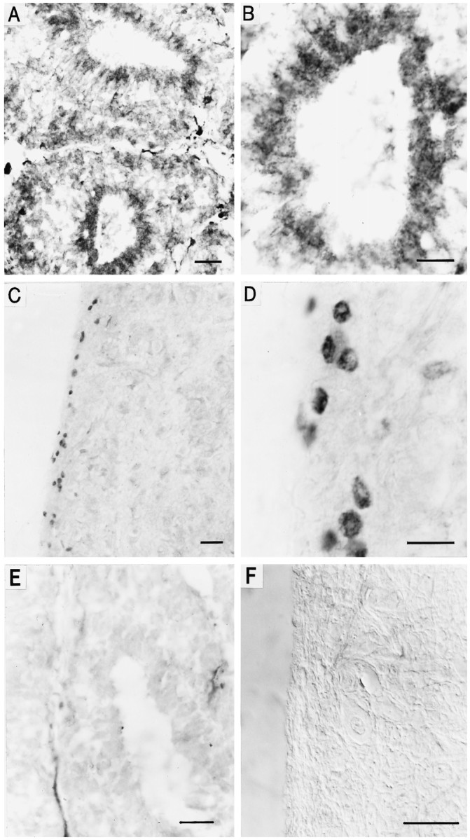
Fig. 3.

Immunohistochemical localization of cone CNG channel in chick and bovine pineal organ. A, Horizontal cryostat section (12 μm) of the chick pineal organ stained with antibody 63–4 and visualized by peroxidase reaction. Cells of two pineal follicles are labeled, whereas pinealocytes outside the follicle are stained less prominently. Scale bar, 20 μm. B, Same as in A at higher magnification. Scale bar, 10 μm. C, Horizontal paraffin section (6 μm) of the bovine pineal organ stained with antibody PPc15 and visualized by peroxidase reaction. PPc15 immunoreactivity is observed in only a few cells in the cortex of the pineal, whereas cells of the medullary part of the organ are not labeled. Scale bar, 20 μm. D, Same as in C at higher magnification. Scale bar, 10 μm. E, Control section of chick pineal. Specific staining is abolished after preincubation of antibody 63–4 with the respective antigenic peptide (20 μg/ml). Scale bar, 20 μm.F, Control section of bovine pineal. No staining was observed when the primary antibody PPc15 was omitted. Picture was taken with Nomarski optics. Scale bar, 20 μm.
