
Fig. 1.
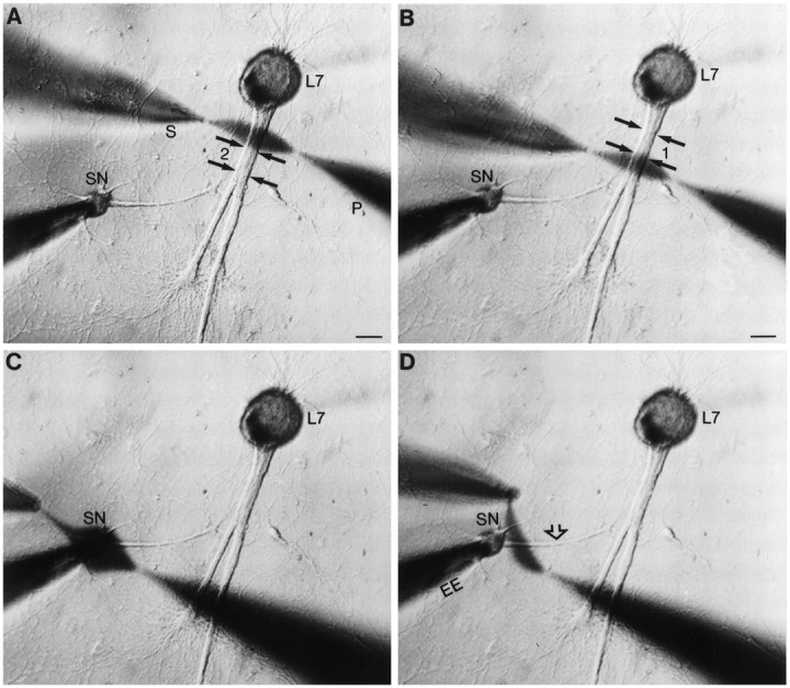
Focal application of neuromodulator to different portions of SN. A–D, Low-power phase-contrast micrographs of 4 d SN–L7 culture. Micropipettes for pressure injection (P) and rapid suction (S) of injected solutions are placed opposite a given region. The pressure and vacuum strengths needed to produce a stream of a given width are determined for each pair of pipettes before placement by the cells. The major axons of L7 emerge from the L7 cell body and extend toward the bottom of the micrographs (double black arrows in A andB). The axon of the SN (open arrow in D) emerges from the SN cell body and extends toward the motor axons. The location of regenerated SN neurites and varicosities in contact with the motor axons is determined with epifluorescent microscopy after dye injections (see Figs. 2, 3). The extracellular electrode near the SN cell body is used to depolarize the cell to evoke an action potential. Intracellular electrode in L7 cell body (out-of-focus shadow extending in the top right of micrographs) is used to record EPSPs. A,B, Examples of focal streams of ∼50 μm across two nonoverlapping regions of the motor axon. Zone 1contained a high density of SN varicosities, whereas zone2 contained proximal SN neurites with few varicosities. Scale bars, 50 μm. C, D, Examples of focal streams across the SN cell body (C) and adjacent region of SN axon (D).