
Fig. 1.
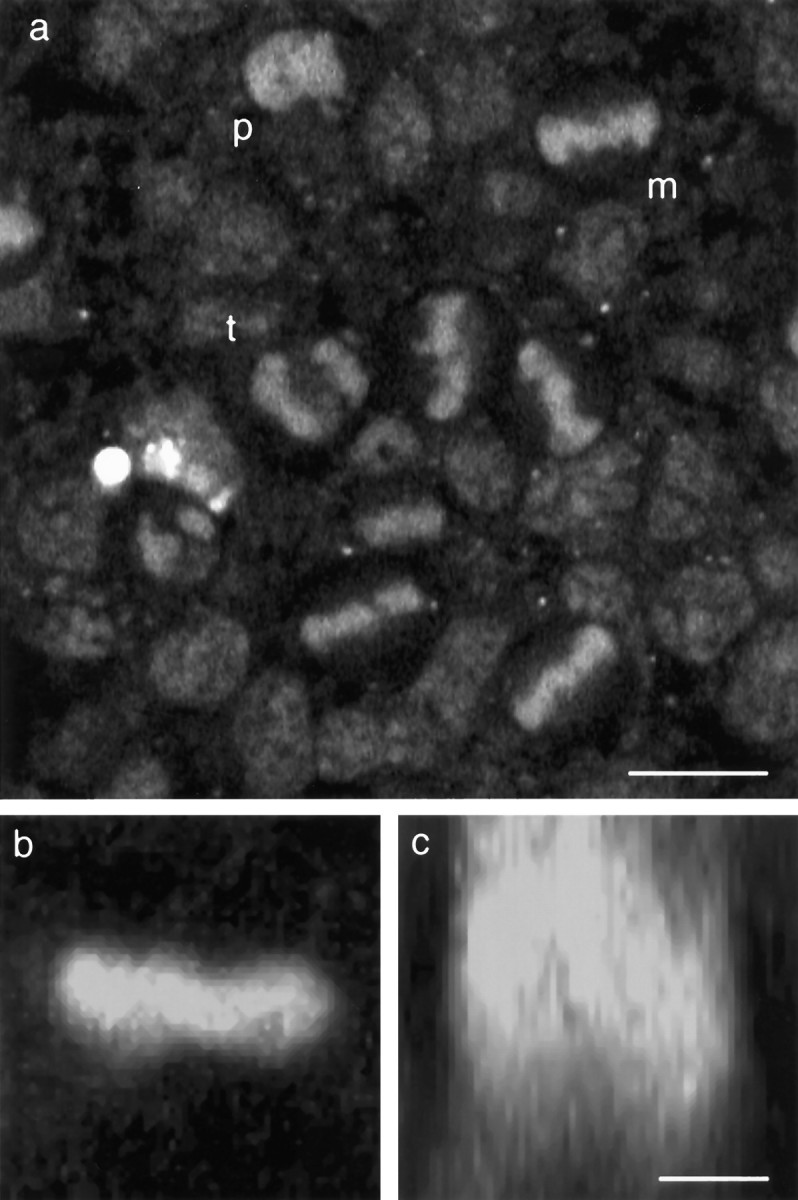
Confocal microscopy of mitotic cells at the ventricular surface of living neuroepithelium from the rat cerebral cortex. Explants from the lateral wall of the neocortical neuroepithelium of embryonic rat between the ages of E12 and E20 were cultured in vitro. The chromatin dye acridine orange (3.3 μm) was used to visualize mitotic figures at the ventricular surface of the tissue imaged by confocal microscopy.a, Single optical section taken parallel to the surface of a sheet of tissue from an E14 rat embryo. Mitotic nuclei in prophase (p), metaphase (m), and anaphase/telophase (t) can be seen. b,c, Two-dimensional extended-focus images generated from thirty-two 1 μm optical sections through a single metaphase cell dividing parallel to the surface of the epithelium. Projected along the apico–basal axis, the image appears as a bright bar (b) but rotated 90° to project along the axis of the spindle shows this to be a disk of chromatin within the depth of the tissue (c). Scale bars: a, 10 μm;b, c, 5 μm.