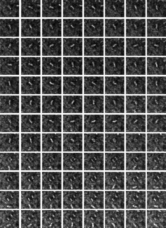
Fig. 2.

Time-lapse sequence of a single mitotic cell in the neuroepithelium of an E19 rat embryo. Neuroepithelium from an E19 embryo was stained with acridine orange and then imaged at 30 sec intervals for 1 hr by time-lapse confocal microscopy. Frames proceed by rows from left to right starting at thetop left. The metaphase plate of the cell seen in the center of frame 1 is in constant motion until frame 61 (row 8, column 5), 30 min into the sequence, when it enters anaphase. The two sets of daughter chromatids separate over the remainder of the frames with little change in the orientation of the spindle. Each frame is 40 μm wide.
