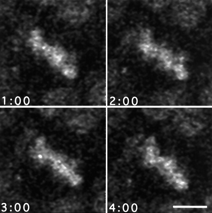
Fig. 3.
Chromosomal movements within the metaphase plate during rotation. Four frames from a sequence of high-magnification images taken at 60 sec intervals during metaphase showing the changes in shape of the metaphase plate, consistent with the oscillatory behaviors of individual chromosomes described by others (Rieder et al., 1994). These movements are superimposed on the larger scale rotation of the entire spindle within the dividing cell. Scale bar, 5 μm.

