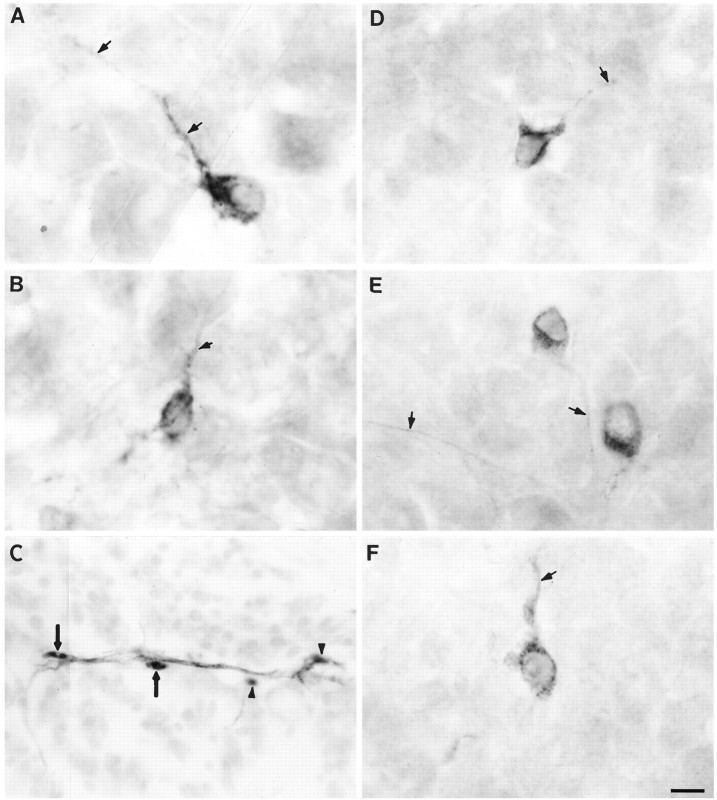
Fig. 3.
Phenotypes of transplanted cells within host embryos. A and B show transplanted dorsal cells stained with QN (quail-specific neuron-specific Ab) that differentiated into peripheral neurons within the host DRG (arrows point to stained neurites). C, Transplanted donor cells (black-stained with Q/CPN) associated with the branch of a small muscle nerve (gray-stained with TuJ1). Two transplanted dorsal cells (arrows) have the morphology of Schwann cells; the other two are out of focus (arrowheads).D–F show transplanted younger ventral cells (stained with QN Ab) within the host DRG that also differentiated into neurons. Arrows point to stained neurites. Scale bar, 10 μm.

