
Fig. 4.
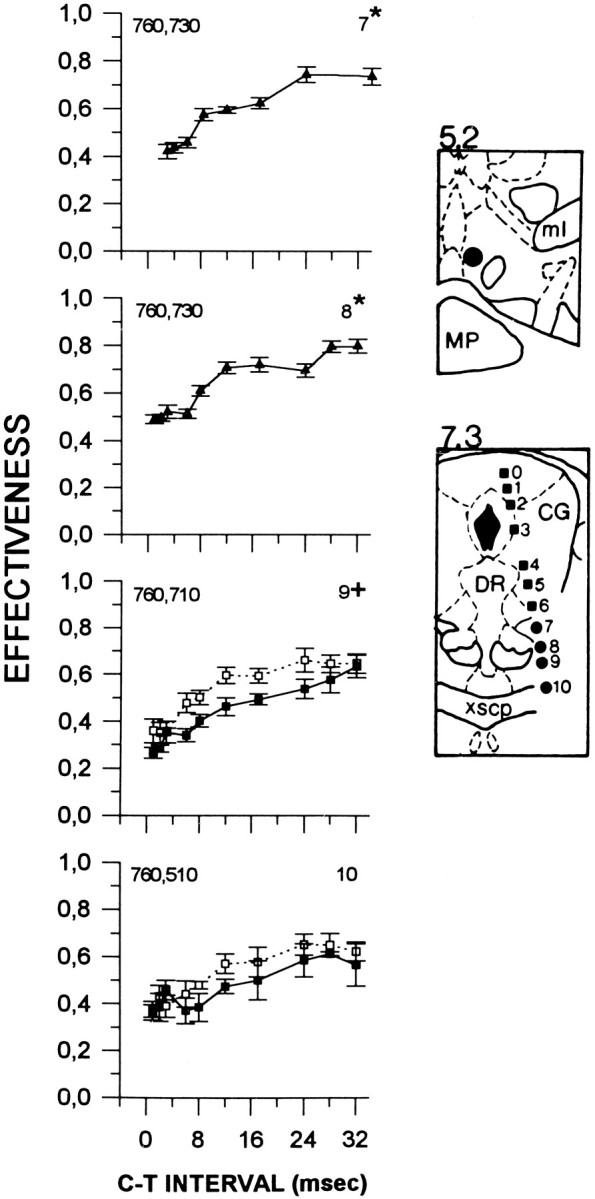
Collision data from subject F9. Locations of the anterior and posterior stimulation sites are illustrated in thetop and bottom panels, respectively. See Figure 1for details. ml, Medial lemniscus; MP, medial posterior mammillary nucleus; CG, central gray;DR, dorsal raphe; xscp, decussation of the superior cerebellar peduncle.