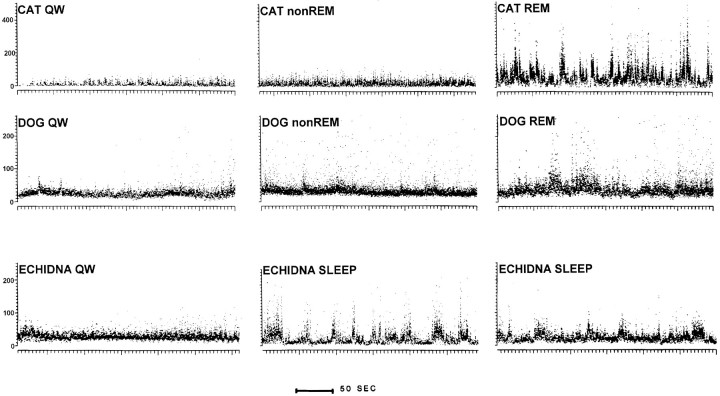
Fig. 4.
Instantaneous compressed rate plots of representative units recorded in nucleus reticularis pontis oralis of the cat, dog, and echidna. Each point represents the discharge rate for the previous interspike interval. In cat QW and non-REM sleep, the discharge rate is low and relatively regular. The rate increases and becomes highly variable during REM sleep. A similar pattern can be seen in a unit recorded in the dog. In the echidna, sleep is characterized by variable unit discharge rates.

