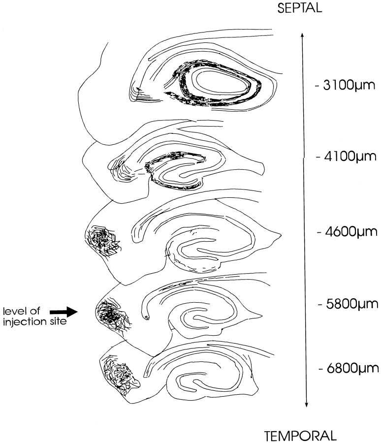
Fig. 7.
Schematic drawing of the topographical organization of the “classical” entorhino-dentate projection originating from entorhinal cell layers II–III. This drawing illustrates the septo-temporal distribution of entorhino-dentate fibers after a PHAL deposit into layers I–III of the entorhinal area. Camera lucida drawings of five septo-temporal levels are shown (vertical levels given in micrometers from bregma) (Paxinos and Watson, 1986). Almost no fibers can be seen in the dentate gyrus at the level of the PHAL injection site (arrow) (septo-temporal extent of the injection site: 500–800 μm). Entorhino-dentate fibers originating in layers I–III follow a rostral trajectory and terminate near the septal pole of the dentate gyrus.

