
Fig. 5.
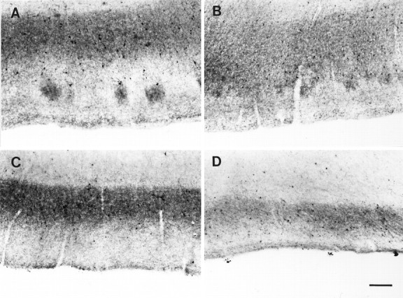
Parvalbumin immunostaining in AD cases.A–D, Distribution of parvalbumin immunostaining at pathological levels 1–4, respectively. Note the eventual complete disappearance of immunolabel in layer II and the consequent reduction on the size of the band of immunoreactivity in layer IIIb. Scale bar, 250 μm.