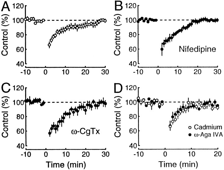
Fig. 4.
Dynorphin release is not associated with a specific presynaptic Ca2+ channel subtype. Two independent mossy fiber pathways were monitored. After baseline responses were stable for at least 10 min, a tetanus was given in one pathway at time 0. The mossy fiber field potentials of the untetanized pathway are plotted against time in normal Ringer’s solution (n = 11; A), 30 μmnifedipine (n = 5; B), and 1 μm ω-CgTx (n = 6;C). The same was done for 1 μm ω-Aga IVA (n = 4; D, filled circles) and 10 μm CdCl2(n = 5; D, open circles); however, as in Figure 2, C andD, a brief 25 Hz train was used to restore responses, and the amplitude of the responses to the last pulse was measured. These results indicate that neither the release nor the inhibitory effects of dynorphin depend on L-, N-, or P-type Ca2+channels.

