
Fig. 4.
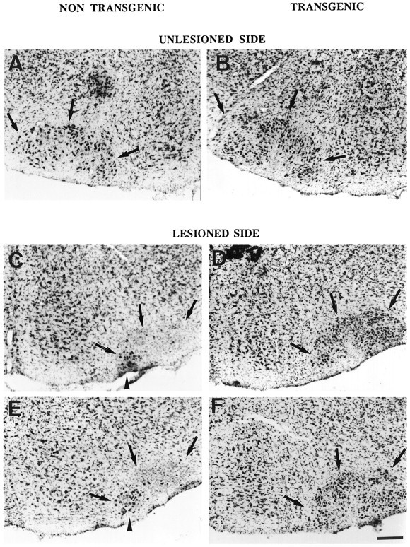
Photomicrographs showing the facial nuclei (dark arrows) in representative 9-d-old animals of each of the four groups after transection of the right facial nerve. Unlesioned side: (A) wild type, (B) wild type–transgenic. Lesioned side: (C) wild type, (D) wild type–transgenic, (E)wobbler, (F) WAT. The black arrowhead points to the ventromedial facial motoneurons, the axons of which are preserved by the transection. Scale bar, 180 μm.