
Fig. 5.
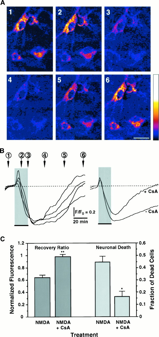
Blockade of the PTP enhances recovery of the NMDA-induced collapse of ΔΨ and decreases neuronal death.A, Digital images of TMRE fluorescence of neurons exposed for 20 min to 200 μm NMDA in the presence of 1.6 μm CsA. Numbers indicate critical time points in the ΔΨ signal shown in B and code panels1 to 6 that are pseudocolored images of TMRE fluorescence. Neurons show a marked depolarization of ΔΨ evoked by the NMDA pulse; repolarization occurs after removal of the stimulus. Scale bar, 20 μm. All other conditions are as in Figure 4.B, Left panel, Time course of ΔΨ from neurons shown in A, measured in arbitrary fluorescence units. Each trace corresponds to one of the neurons in the digital images. Right panel, Comparison of the time courses of ΔΨ in response to 20 min NMDA in the presence or absence of CsA. Traces are averages from all recordings (+CsA:n = 26; −CsA: n = 74). C, Recovery of ΔΨ (Recovery Ratio) was calculated for recordings obtained in the presence of CsA (n = 26 neurons) and compared with those presented in Figure 4C for 20 min NMDA. Peak depolarization is not affected by CsA (Peak Depolarization = 0.351 ± 0.048; p = 0.1). Cell death induced by NMDA was simultaneously assessed in the presence (n = 7126 neurons) and absence (n = 7985 neurons) of CsA for six independent experiments. Fraction of dead cells in cultures treated with vehicle was subtracted (0.054 ± 0.003; n = 6547 neurons). Bars represent mean ± SEM. *, Statistically significant difference between treatments (p < 0.002); **p < 0.0001.