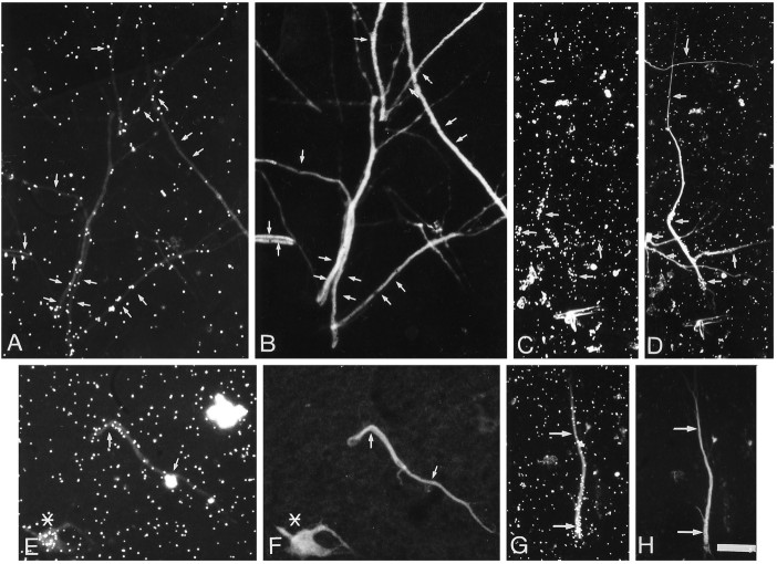
Fig. 3.
Galactose and fucose incorporation in isolated dendrites. Neurites isolated from 10- to 12-d-old cultures were preincubated for 1 hr in low-glucose medium, pulse-labeled with 200 μCi/ml [3H]fucose or 100 μCi/ml [3H]galactose for 1 hr, rinsed in medium containing 10 mm cold sugar, and fixed and immunostained for MAP2.A, C, Autoradiographic localization of the sites of [3H]galactose incorporation.E, G, Autoradiographic localization of [3H]fucose incorporation. B,D, F, H, The parts show that the sugar incorporation is localized over MAP2-stained processes. The arrows indicate the localization of dendrites identified by MAP2 staining. The asterisk indicates the presence of a labeled neuron that was identified by the DNA staining. Scale bar, 25 μm.

