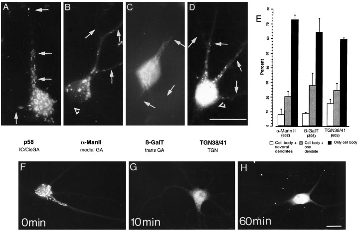
Fig. 8.
Dendritic localization of different Golgi compartments in hippocampal neurons in culture. Fourteen-day-old cultures were stained for p58 (A), α-ManII (B), β-galactosyltransferase (βGalT) (C), or TGN 38/41 (D). Stained puncta were observed concentrated within the cell body and frequently extending into dendrites that were identified by double staining for MAP2 (arrows). The staining was not found in axons (open triangles). E, Distribution of different Golgi compartments in 14- to 20-d-old hippocampal neurons in culture. Stained cells were counted and classified according to whether the immunostained structures were localized only in the cell body, in the cell body and one dendrite, or in the cell body and several dendrites. The bars represent the percentage ± SEM of cells having a given staining distribution. Thenumbers in parentheses represent the number of cells counted for each antibody tested. The lower section shows the effect of brefeldin A on the localization of Golgi organelles. Fifteen-day-old neurons were stained for α-mannosidase II after treatment with 5 μg/ml brefeldin A. F, Control; G, 10 min in brefeldin A;H, 60 min in brefeldin A. Note the rapid redistribution of this marker induced by brefeldin A. Scale bars, 25 μm.

